
Product Overview
Gamma-aminobutyric acid (GABA) is the main inhibitory neurotransmitter of the central nervous system (CNS) and participates in 40% of the inhibitory synapses of adult vertebrates [1]. There are several types of GABAergic neurons in the brain, differing in the areas they are formed and where they migrate after neurogenesis [2].
Here, we provide cortical GABAergic interneurons, which mediate neuronal function by interacting with excitatory glutamatergic neurons. GABA signaling has been involved in a plethora of CNS disorders such as Autism [3], Parkinson’ Disease [4], and Huntington’s Disease, among others.
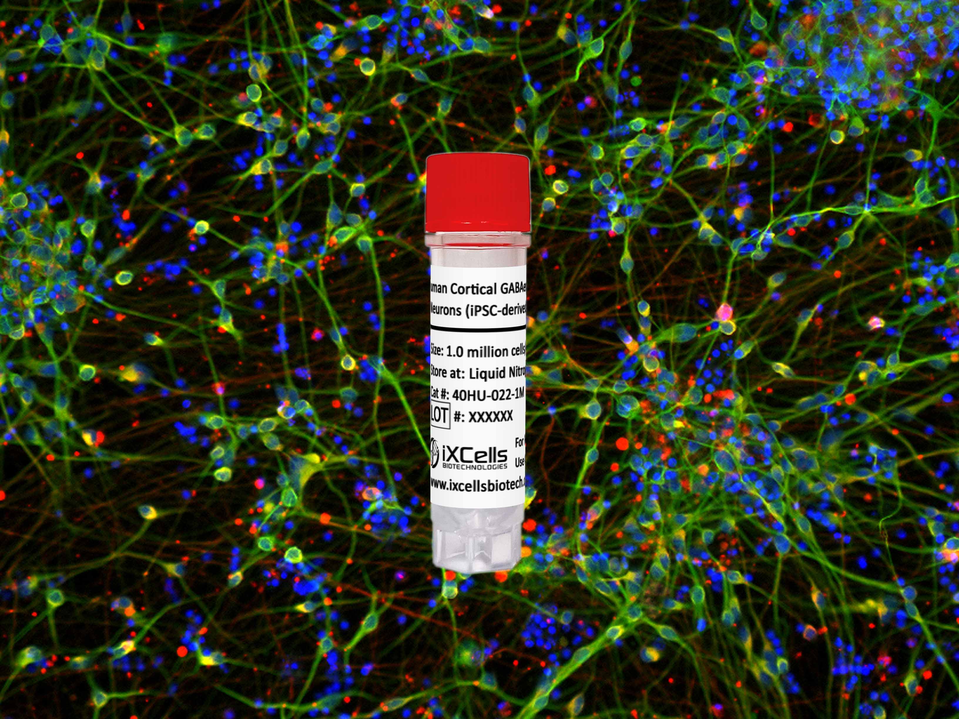
iXCells Biotechnologies is proud to provide fully differentiated and functional Human Cortical GABAergic Neurons 2.0 (iPSC-derived) which display typical neuronal morphology (Figure 1), and express typical markers of GABAergic neurons, e.g. GAD67, vGAT (Figure 2, 3), when cultured in the GABAergic Neuron Culture Medium (Cat# MD-0122-100ML). Gamma-aminobutyric acid (GABA) release could be measured by ELISA after 7 days maturation (Figure 4). Moreover, Human GABAergic Neurons 2.0 present spontaneous neuron activity when co-cultured with astrocytes (Figure 5), as assessed by Multi-Electrode Array (MEA), and this activity increases with maturation.

Figure 1. Cells were seeded in Poly-D-lysine/Laminin coated plates at a density of 150,000 neurons/cm2. Representative bright field images of GABAergic neurons 2.0 at different time points after thawing.

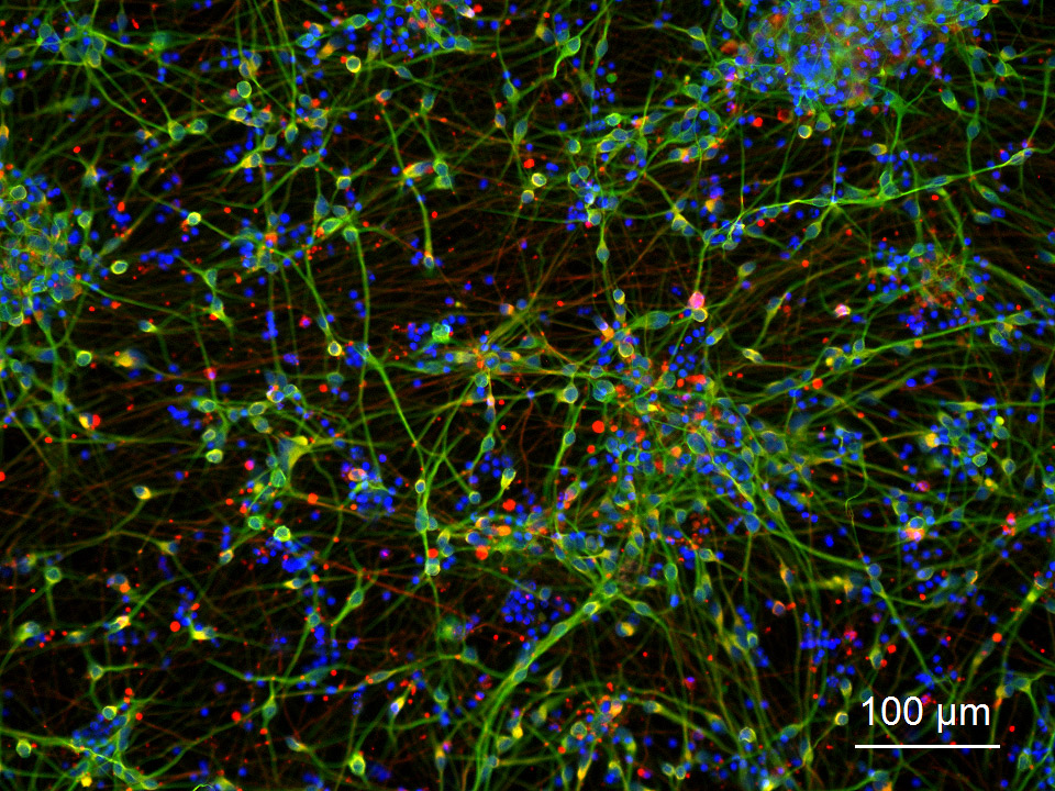
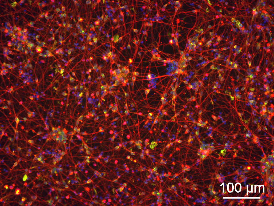
Figure 2.Cells were recovered and seeded in Poly-D-lysine/Laminin coated plates at a density of 150,000 neurons/cm2 for 7 days. Immunofluorescence staining showing GAD67/MAP2 (A) and VGAT/Tuj1 (B) positive cells on day 7 in culture. DAPI staining (blue) is also shown. Scale bar: 100µm.
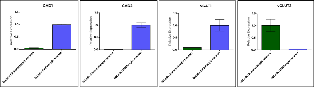
Figure 3. Quantification of RNA expression of the indicated genes by quantitative PCR. Cells were seeded at a density of 150,000 cells/cm2 in Poly-D-Lysine/Laminin coated plates and cultured for 14 days post-thawing, then harvested and RNA was extracted for quantification. GAD1, GAD2 and vGAT1 are GABAergic neuron marker genes, and vGLUT2 is a glutamatergic neuron marker gene.

Figure 4. Quantification of GABA in cell culture media by ELISA (Immusmol, Cat#BA-E-2500). Cells were seeded at a density of 100,000 cells/cm2 in Poly-D-Lysine/Laminin coated plates and cultured for 7 days. Media samples were collected after 72h of culture post the last media change. GABA ELISA was performed following manufacturer’s instructions. One-way ANOVA was used to determine statistical significance (***p<0.001), against iPSC control.
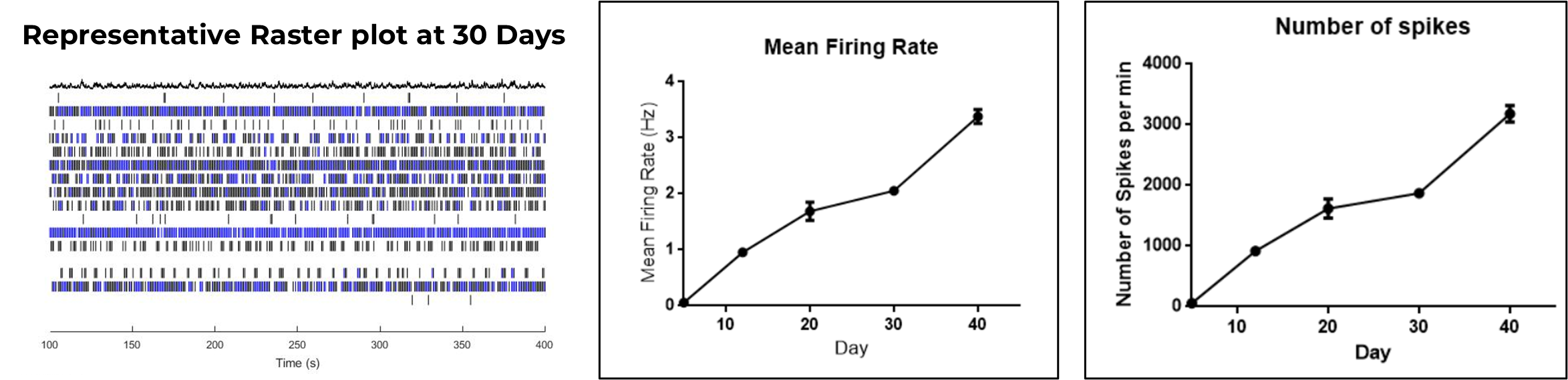
Figure 5. Quantification of Multi-Electrode-Array (MEA) metrics. Cells were co-cultured with astrocytes in 24-well MEA plates, at a density of 100,000 neurons/well and 50,000 astrocytes/well in Polyethyleneimine/Laminin coating. A raster plot showing neuronal network activity and burst for each electrode is shown on the left panel. Mean firing rate quantification is shown on middle panel. Total number of spikes quantification is shown on right panel. The data was generated using Axion Biosystems analysis software.
Product Details
| Organism | Homo Sapiens, Human |
| Cell Type | Brain Cell |
| Tissue | N/A |
| Disease | N/A |
| Package Size | 1 x 10⁶ cells/vial, 2 x 10⁶ cells/vial |
| Passage Number | P0 |
| Growth Properties | Adherent |
| Product Format/Shipped | Cryopreserved |
| Storage | Liquid Nitrogen |
| Associated Media | GABAergic Neuron Culture Medium (MD-0122) |
References
[1] Nicholas CR, Chen J, Tang Y, et al. Functional maturation of hPSC-derived forebrain interneurons requires an extended timeline and mimics human neural development. Cell Stem Cell. 2013.
[2] Huang ZJ, Paul A. The diversity of GABAergic neurons and neural communication elements. Nat Rev Neurosci. 2019.
[3] Zhao H, Mao X, Zhu C, et al. GABAergic System Dysfunction in Autism Spectrum Disorders. Front Cell Dev Biol. 2022.
[4] Murueta-Goyena A, Andikoetxea A, Gómez-Esteban JC, Gabilondo I. Contribution of the GABAergic System to Non-Motor Manifestations in Premotor and Early Stages of Parkinson’s Disease. Front Pharmacol. 2019.