


Product Overview
Sensory neurons play a crucial role in the detection and response to different types of sensory stimuli, such as touch, temperature, and pain. As one of the most significant neuronal subtypes in the human peripheral nervous system, they form neuronal-glial networks that are responsible for a variety of motor and sensory mediated functions [1]. Dysfunction of sensory neurons can lead to various neurological disorders such as pain, multiple sclerosis (MS), amyotrophic lateral sclerosis (ALS), and problems with mechano- or temperature perception [2]. Understanding the biology of iPSC-derived sensory neurons can help with new treatments for these disorders, as well as improve our understanding of normal sensory function [3].
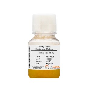
iXCells Biotechnologies is proud to provide fully differentiated and functional human iPSC-derived human sensory neurons that display typical neuronal morphology and express key markers e.g., Peripherin, BRN3A (Figure 1) when cultured in the Human Sensory Neuron Maintenance Medium (Cat# MD-0114-100ML). In addition, our iPSC-derived sensory neurons can also be co-cultured with glia or other cell types for drug screening platforms.
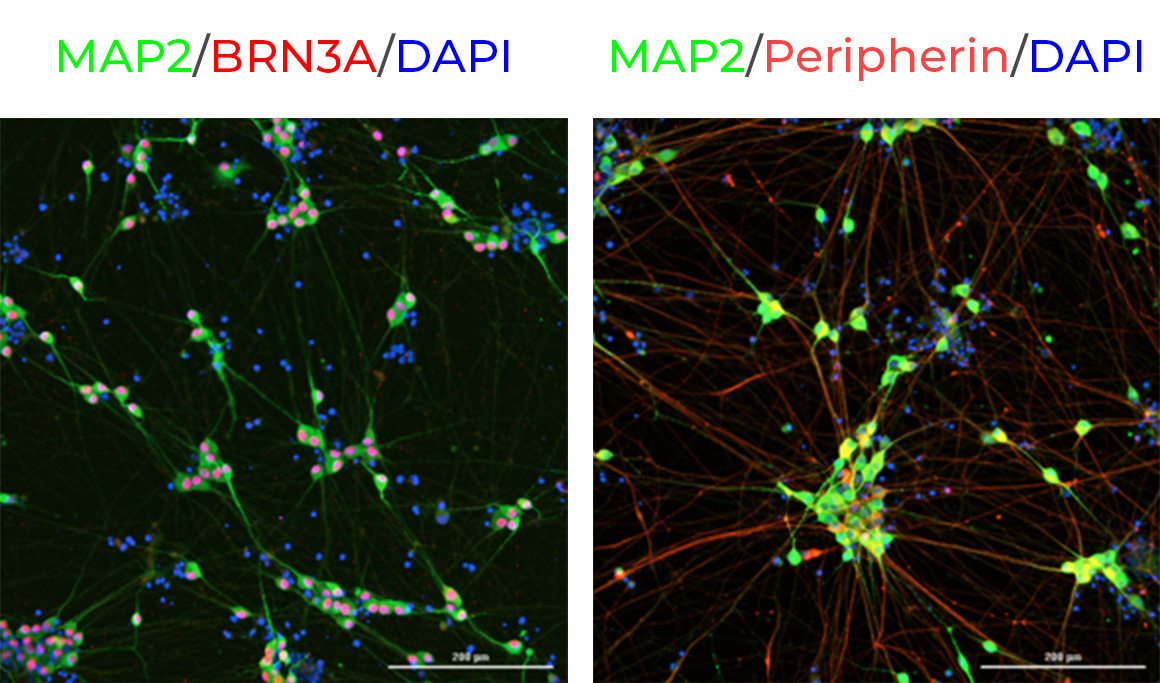
Figure 1. Immunostaining of iPSC-derived sensory neurons expressing MAP2 (green), BRN3A (red), Peripherin (red) 7 days post-thaw. All cells were counterstained for DAPI (blue). Scale bars: 200 µm.
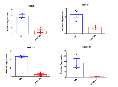
Figure 2. Gene expression values for NTRK1 (TRKA), TRPV1, SCN9A (Nav1.7) and SCN10A (Nav1.8) from 3 weeks post-thaw sensory neurons and control (iPSC-derived Astrocytes). Results are graphed as mean ± SEMs.
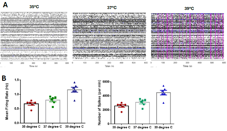
Figure 3. (A) Example raster plot showing spikes (black lines) and network bursts (pink lines) at different temperatures in the MEA for sensory neurons grown for 2 weeks. (B) The electrical parameters were measured in the MEA over different temperatures and the results are graphed as mean ± SEM for mean firing rate.
Product Details
| Organism | Homo Sapiens, Human |
| Cell Type | Sensory Neuron |
| Tissue | Human Brain |
| Disease | Normal |
| Package Size | 1 x 10⁶ cells/vial, 2 x 10⁶ cells/vial |
| Passage Number | P0 |
| Growth Properties | Adherent |
| Product Format/Shipped | Cryopreserved |
| Storage | Liquid Nitrogen |
| Associated Media | Human Sensory Neuron Maintenance Medium (Cat# MD-0114-100ML) Recovery Supplement (Cat# MD-0110-20μL) Y27632 (Cat# MD-0025-2MG) |
References
[1] Basbaum AI, Bautista DM, Scherrer G, Julius D. Cellular and molecular mechanisms of pain. Cell. 2009 16;139: 267-84.
[2] Fargeot G, Echaniz-Laguna A. Sensory neuronopathies: new genes, new antibodies and new concepts. J Neurol Neurosurg Psychiatry. 2021 9:jnnp-2020-325536
[3] Lampert A, Bennett DL, McDermott LA, Neureiter A, Eberhardt E, Winner B, Zenke M. Human sensory neurons derived from pluripotent stem cells for disease modelling and personalized medicine. Neurobiol Pain. 2020 18;8:100055.
