Product Description
Schwann cells are neural crest derivatives that ensheathe and myelinate axons of peripheral nerves [1]. Each Schwann cell wraps around the shaft of an individual peripheral axon, forming myelin sheaths along segments of the axon. Schwann cells play important roles in the development, function, and regeneration of peripheral nerves. When an axon is dying, the Schwann cells surrounding it aid in its digestion, leaving an empty channel formed by successive Schwann cells, through which a new axon may then grow from a severed end. The number of Schwann cells in peripheral nerves is tightly regulated [2]. Their proliferation in vitro can be stimulated by various growth factors including PDGF, FGF, neuregulin, and others [3]. Schwann cells provide a relatively simple, well-defined, and accessible mammalian model for the study of a number of neuronal developmental questions.
Product Details
| Organism | Mus musculus, Mouse |
| Cell Type | Schwann Cells |
| Tissue | Sciatic nerve |
| Disease | Normal |
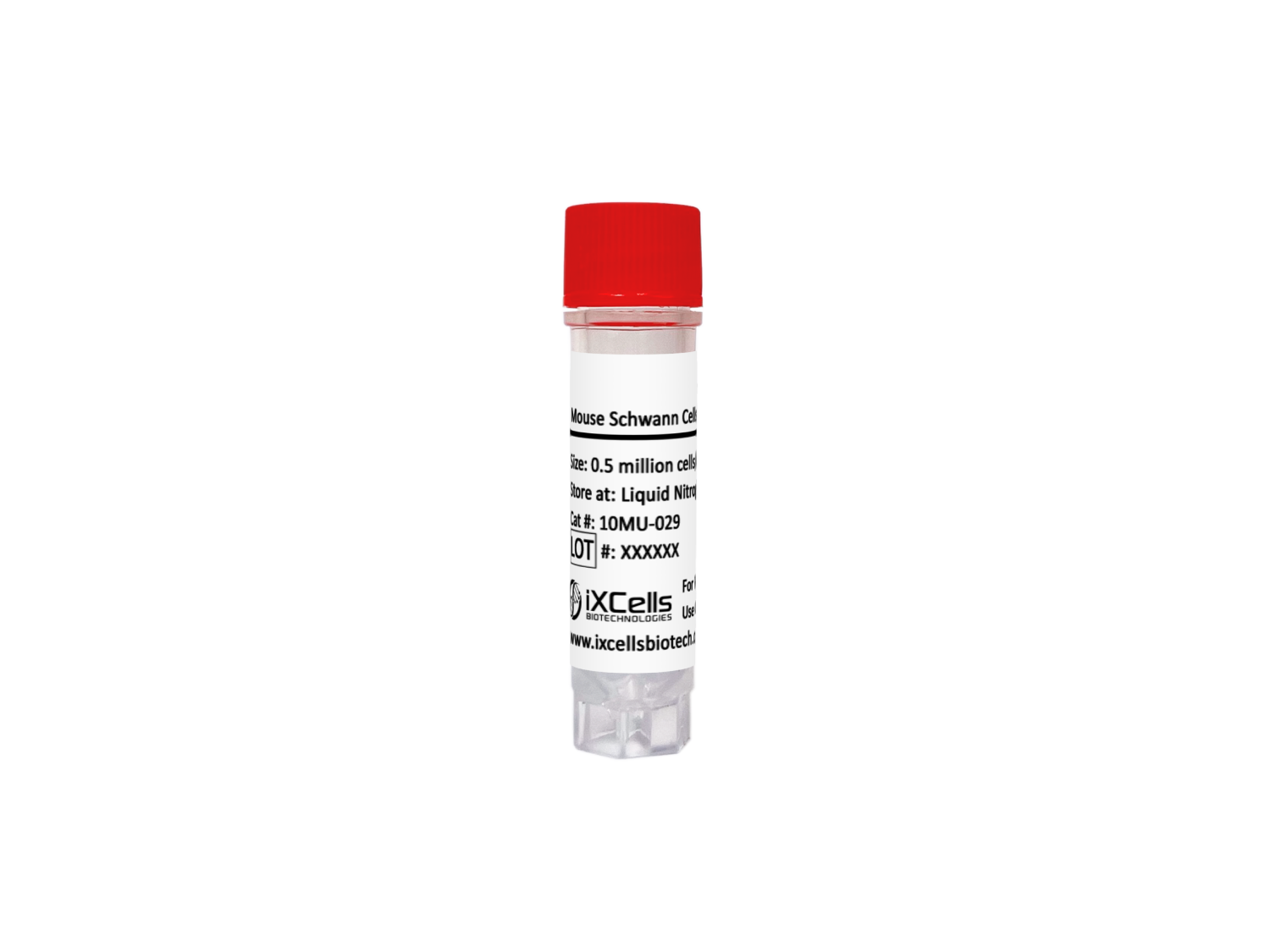
| Package Size | 0.5 x 10⁶ cells/vial |
| Passage Number | P1 |
| Growth Properties | Adherent |
| Product Format/Shipped | Cryopreserved |
| Storage | Liquid Nitrogen |
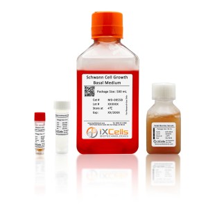
| Associated Media | Schwann Cell Growth Medium (Cat# MD-0055) |
iXCells Biotechnologies provides high quality Mouse Schwann Cells (MSC), which are isolated from mouse sciatic nerves and cryopreserved at P1, with >0.5 million cells in each vial. MSC express S-100. They are negative for HIV-1, HBV, HCV, mycoplasma, bacteria, yeast, and fungi and can further expand in Schwann Cell Growth Medium Schwann Cell Growth Medium (Cat# MD-0055) for two additional passages under the condition suggested by iXCells Biotechnologies.
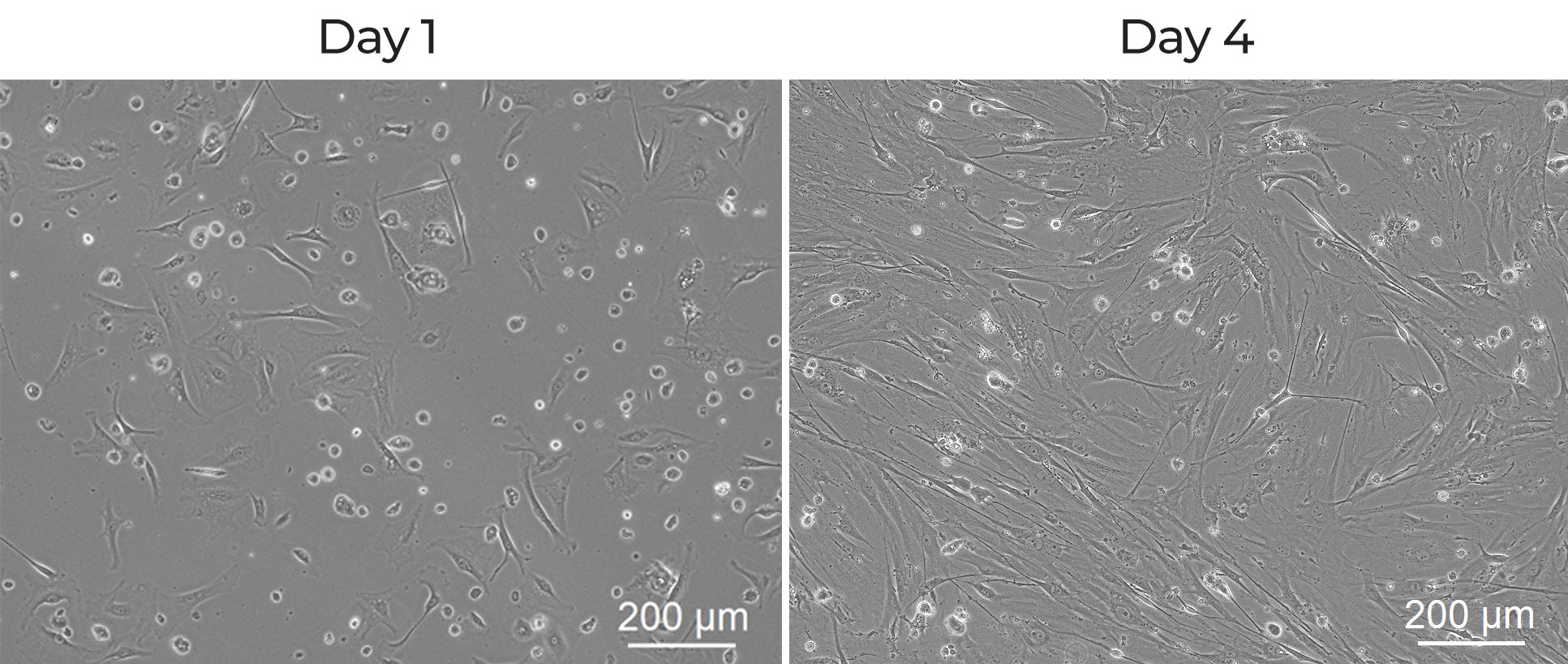
Figure 1. Mouse Schwann Cells (MSC). The cells were recovered, and seeded at 10,000 cells/cm2 following iXCells protocol. Phase contrast images were taken at the indicated time post recovery.
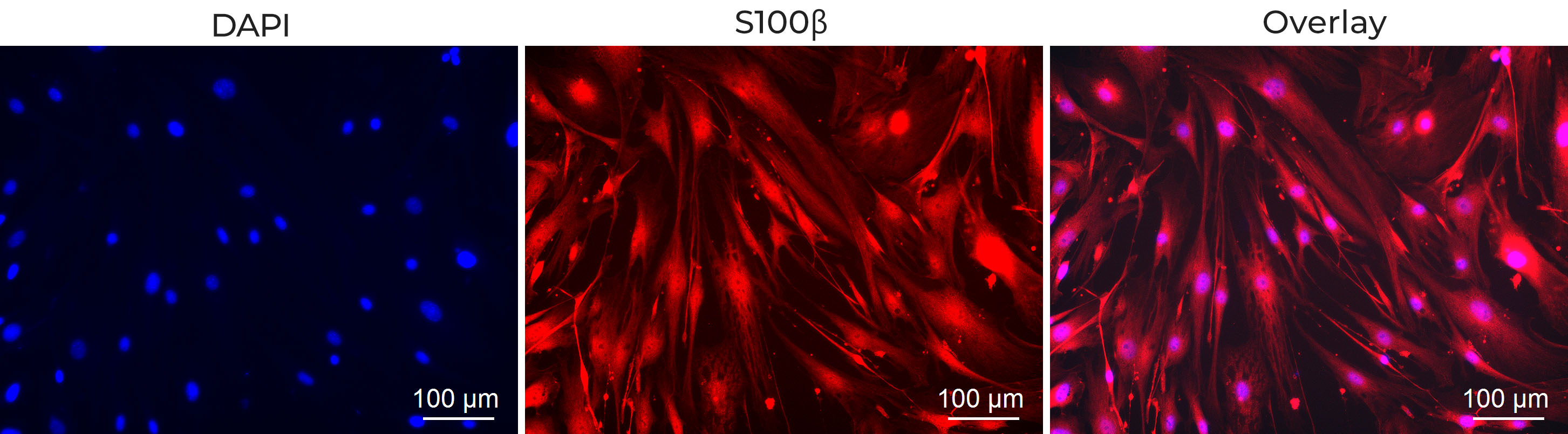
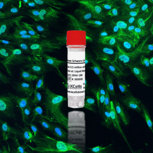
Figure 2. Immunofluorescence staining of MSC with the antibody against S100β (Red). Nuclei were counterstained by DAPI (Blue).
References
[1] Negro S., Pirazzini M., Rigoni M. (2022). Models and methods to study Schwann cells. J Anat. 2022 Nov;241(5):1235-1258. doi: 10.1111/joa.13606. Epub 2022 Jan 5.
[2] Yang, C., Hawkins, K. E., Doré, S., & Candelario-Jalil, E. (2019). Neuroinflammatory mechanisms of blood-brain barrier damage in ischemic stroke. American journal of physiology. Cell physiology, 316(2), C135–C153.
[3] Godinho-Pereira, J., Garcia, A. R., Figueira, I., Malhó, R., & Brito, M. A. (2021). Behind Brain Metastases Formation: Cellular and Molecular Alterations and Blood-Brain Barrier Disruption. International journal of molecular sciences, 22(13), 7057.