
Product Description
Glomerular podocytes in kidney are highly specialized visceral epithelial cells with a complex cytoarchitecture. These cells feature finger-like projections called “foot process” that interlock with each other, creating tiny gaps known as filtration slits that play a crucial role in filtering blood within the glomerulus [1,2]. Podocytes are involved in a variety of glomerular functions, including glomerular basement membrane turnover, maintenance of filtration barrier, support of the capillary tuft, regulation of glomerular filtration and immunological functions. Injury to podocytes may lead to proteinuria, a hallmark of most glomerular diseases and chronic kidney disease. Disruptions of podocyte architecture resulting in the retraction of foot processes and proteinuria are common features in the progression of acquired glomerular disease [3,4].
iXCells Biotechnologies offers high quality primary Human Podocytes isolated from human kidney obtained by following IRB protocol and cryopreserved at P2, with ≥ 0.5 million viable cells in each vial. These podocytes have been characterized by the proliferation, and the expression of cell type specific markers, including CD2AP, Nephrin, NPHS2, Podocin, Synaptopodin (Figure 1, 2). Human Podocytes are negative for HIV-1, HBV, HCV, mycoplasma, bacteria, yeast, and fungi. These cells can be maintained in Podocyte Culture Medium (Cat# MD-0120) and expanded for 2~3 passages under conditions suggested by iXCells Biotechnologies. Further expansion may decrease podocyte particular traits such as foot processes and expression of slit diaphragm proteins.

Figure 1.Human Podocytes. The cells were recovered, and seeded at 10,000 cells/cm2 following iXCells’ protocol. Phase contrast images were taken at the indicated time post-recovery.
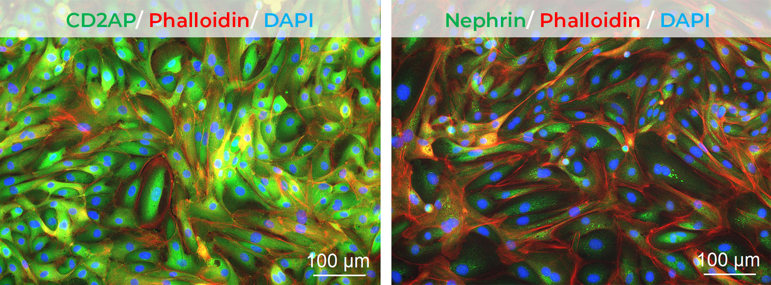

Figure 2. Immunofluorescence staining of Human Podocytes with antibodies against CD2AP, Nephrin, NPHS2, Podocin, Synaptopodin (Green), and Phalloidin (Red), separately. Nuclei were counterstained by DAPI (Blue).
Product Details
| Organism | Homo Sapiens, Human |
| Cell Type | Epithelial Cell |
| Tissue | Human Kidney |
| Disease | Normal |
| Package Size | 0.5 x 10⁶ cells/vial |
| Passage Number | P2 |
| Growth Properties | Adherent |
| Product Format/Shipped | Cryopreserved |
| Storage | Liquid Nitrogen |
| Associated Media | Podocyte Culture Medium (MD-0120) |
References
[1] Pavenstadt H., Kriz W., Kretzler M. Cell biology of the glomerular podocyte. Physiol Rev. 2003;83(1):253–307.
[2] Romagnani P., Remuzzi G., Glassock R. Chronic kidney disease. Nat Rev Dis Primers. 2017;3:17088.
[3] Patrakka J., Tryggvason K. New insights into the role of podocytes in proteinuria. Nat Rev Nephrol. 2009; 5(8):463–468.
[4] Levey A.S., Coresh J. Chronic kidney disease. Lancet. 2012;379(9811):165–180.