



Product Description
Human umbilical vein endothelial cells (HUVEC) are the cells derived from the endothelium of veins from the umbilical cord. They exhibit a cobblestone phenotype when lining vessel walls. HUVECs are commonly used as a laboratory model system for physiological and pharmacological studies, such as macromolecule transport, blood coagulation, angiogenesis, and fibrinolysis [1]. Serving as the classic model system, they have been applied into the study of multiple aspects of endothelial function and disease, including normal or tumor-associated angiogenesis, oxidative stress and inflammation related pathways in endothelium under normal and pathological conditions [2].
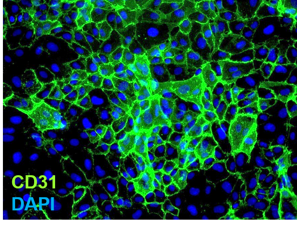
iXCells Biotechnologies provides high quality Human Umbilical Vein Endothelial Cells (HUVEC), which are isolated from human umbilical vein of mix donors, and cryopreserved at P2, with ≥ 0.5 million cells in each vial. HUVEC have “cobblestone” morphology and positive staining with typical endothelial cell markers, including vWF/Factor VIII, VE-Cadherin, and CD31. These HUVEC are negative for HIV-1, HBV, HCV, mycoplasma, bacteria, yeast and other fungi. The cells can further expand no more than 3 passages under the conditions suggested by iXCells Biotechnologies. Additional expansion may decrease the purity.

Figure 1. Human Umbilical Vein Endothelial Cells (HUVEC). The cells were recovered, and seeded at 10,000 cells/cm2 following iXCells’ protocol. Phase contrast images were taken at the indicated time post recovery.
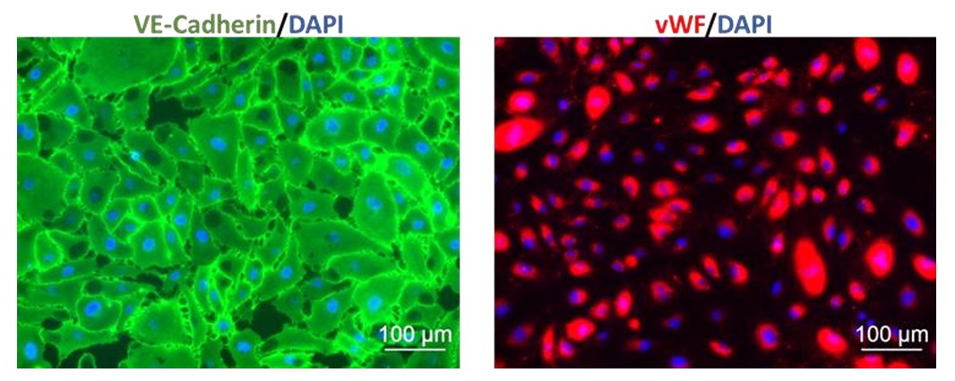
Figure 2. Immunofluorescence staining of HUVEC with antibodies against VE-Cadherin (Green) and vWF (Red). Nuclei were counterstained by DAPI (Blue).
Product Details
| Tissue | Human umbilical cord tissue |
| Package Size | 0.5×10⁶cells/vial |
| Passage Number | P2 |
| Shipped | Cryopreserved |
| Storage | Liquid nitrogen |
| Growth Properties | Adherent |
| Media | Endothelial Cell Growth Medium (Cat# MD-0010) |
References
[1] Park HJ, Zhang Y, Georgescu SP, Johnson KL, Kong D, Galper JB (2006). “Human umbilical vein endothelial cells and human dermal microvascular endothelial cells offer new insights into the relationship between lipid metabolism and angiogenesis”. Stem Cell Rev 2 (2): 93–102.
[2] Nallamshetty S, Chan SY, and Loscalzo J. Hypoxia: a master regulator of microRNA biogenesis and activity. Free Radic Biol Med. 2013; 64: 20-30.
Hunt, R. C., Katneni, U., Yalamanoglu, A., Indig, F. E., Ibla, J. C., & Kimchi‐Sarfaty, C. (2022). Contribution of adamts13 ‐independent vwf regulation in sickle cell disease. Journal of Thrombosis and Haemostasis. https://doi.org/10.1111/jth.15804 -- Learn More
Laiva, A. L., O’Brien, F. J., & Keogh, M. B. (2021). SDF-1α Gene-Activated Collagen Scaffold RESTORES pro-angiogenic wound HEALING features in Human Diabetic Adipose-derived stem cells. Biomedicines, 9(2), 160. doi:10.3390/biomedicines9020160 -- Learn More
Shen, L., Hu, Y., Lou, J., Yin, S., Wang, W., Wang, Y., . . . Wu, W. (2019). CircRNA‑0044073 is upregulated in atherosclerosis and increases the proliferation and invasion of cells by TARGETING MIR‑107. Molecular Medicine Reports. doi:10.3892/mmr.2019.10011 -- Learn More